
In this post we will learn about the External Structures of Bacteria.
In our previous post MORPHOLOGY OF BACTERIA we learnt in detail about the size, shape and forms of bacteria, this post is continuation of the same.
So, let’s get started…
STRUCTURES EXTERNAL TO CELL WALL ARE
- Flagella
- Fimbriae/ Pili
- Glycocalyx/ Capsules
- Sheaths
- Prostheceae & stalks
- Cell wall
We will go through each one of them in detail.
Table of Contents
Toggle1.FLAGELLA
- Bacteria can be motile or non-motile.
- The motile bacteria swim with the help of small flexible, whip like attachment called flagella (singular flagellum).
- Flagellum measure about 120 Å thick and 4-5 µ.
- Chemically they are protein known as Flagellin.
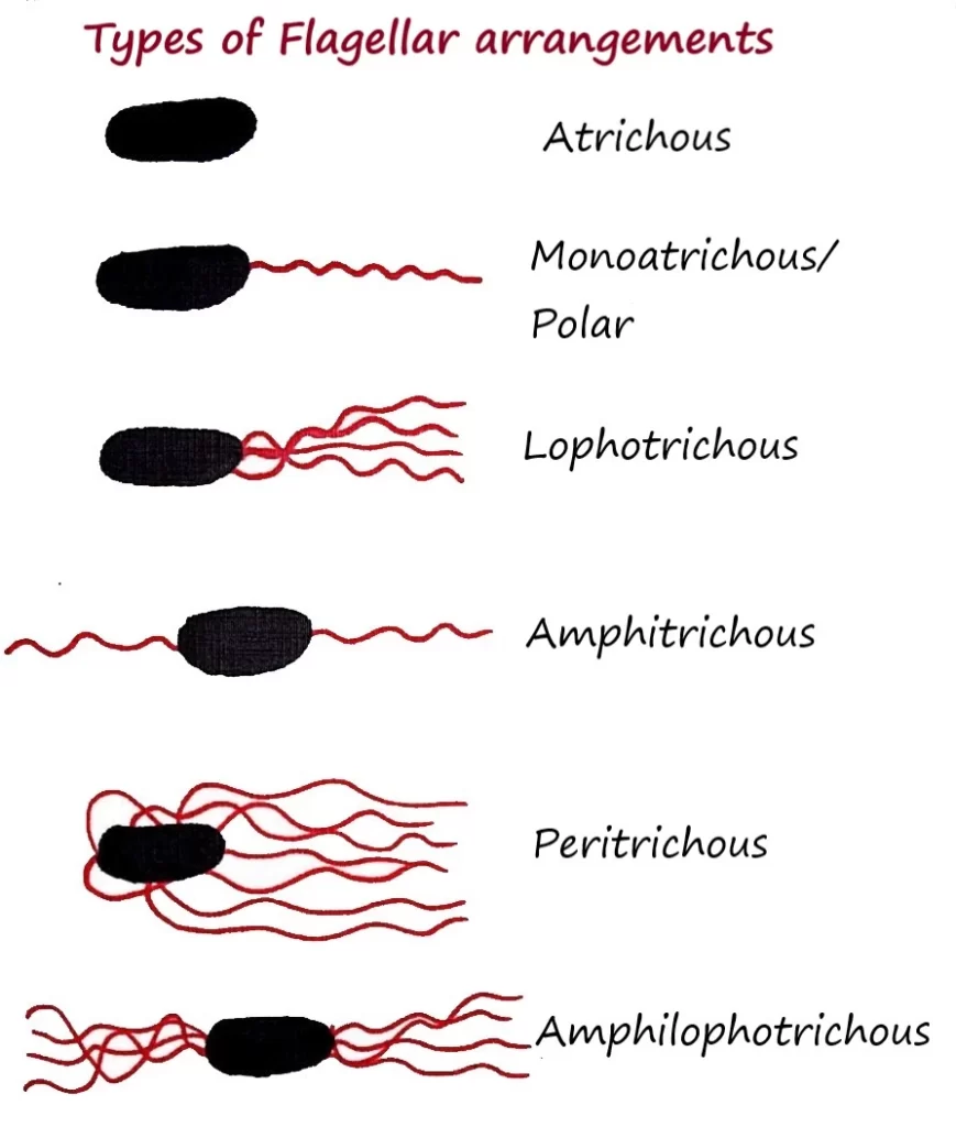
- The bacteria with no flagella are called Atrichous (Diphtheria bacilli).
- Different species of bacteria have variation about number and position of attachment of flagella.
- That is why bacteria are classified into following types:
Monotrichous/ polar: Single flagellum at one pole.
Lophotrichous: Tuft of flagella at one pole.
Amphitrichous: Single flagella at both poles.
Peritrichous: Many flagella attached all over the cell body.
Amphilophotrichous-Tuft of flagella at both ends.
STRUCTURE
- A single flagellum is ten times longer than their bacterial cell.
- A flagellum has 3 main parts:
(1) Basal Body:
- A small rod like structure attached deep to cytoplasm of the cell.
- Cytoplasm provide energy to flagellum.
- Function is to synthesize the polymers of flagellum & regulation of its movement.
(2) Hook
- It arises from cell wall & connects with the basal body and main filament (shaft).
- They provide specific shape to the flagellum.
- Each hook has specific antigenic property.
(3) Filament or Shaft
- It is a tubular structure attached to the hook.
Function
- Provides motility to bacterial cell.
Movement Due to Flagella
- The flagella rotate around the axis.
- The rotation is either clockwise or anti-clockwise.
- They form a bundle which pushes against the surrounding liquid and propels the bacterium.
- When a bacterium moves in specific direction it is called “Run”.
- If a bacterium come across some chemicals & starts to move in non-specific direction it is called “Tumbles”.
- Then it again starts to move in any specific direction.
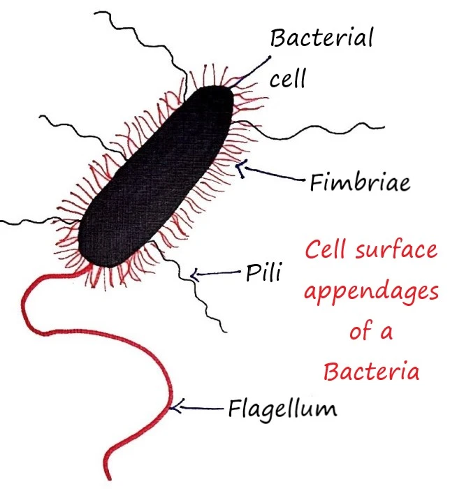
2.FIMBRIAE / PILI
- Many Gram-negative bacteria have thin, short, straight and hair like attachment.
- They contain protein which is known as ‘pilin’.
- They are of two types- fimbriae & pili but their functions are different.
FIMBRIAE
- They have tendency to adhere to each other or to surface.
- They help the bacteria adhere to epithelial surface of body.
- Example- Fimbriae of E. coli helps it to adhere the small intestinal lining.
- If there is no fimbria, colony formation may not happen and no disease occurs.
PILI
- Usually longer than fimbriae.
- They help in motility and transfer of DNA.
- Two types of pili are found in bacteria.
(a) Somatic pili (b) Sex pili or conjugate pili.
(a) Somatic Pili:
Each bacterial cell has about 100 somatic pili which help the bacterium for attachment to a surface.
(b) Sex Pili or Conjugate Pili:
They are also known as F pili and are controlled by sex factors.
Functions of Pili
- They help the bacteria to attach to the surface or to other organism with their adhesive properties.
- They act as an antigen.
- Sex pili transfers the Genetic material from donor to recipient cell.
- They act as bacteriophage receptor.
3.GLYCOCALYX OR CAPSULES
Definition
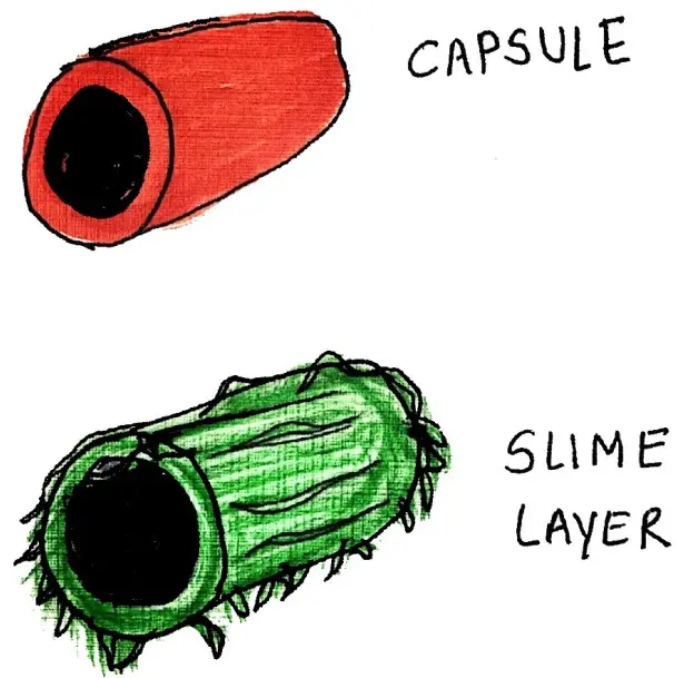
Prokaryotic cell sometime secretes a substance outside the cell wall known as glycocalyx.
- Glycocalyx means the coat of sugar.
- The bacterial glycocalyx is a sticky, gelatinous polymer.
- They are made up of polysaccharide or polypeptide or both.
Capsule
If the substance is organized and is firmly attached to the cell wall, the glycocalyx is described as a capsule.
- They are responsible for disease causing ability of a few types of bacteria.
- It is divided into two groups:
(a) Macrocapsule: It is about O.21 µm thick and can be seen under light microscope.
(b) Microcapsule: It can’t be seen under light microscope but can be demonstrated immunologically.
Slime layer
- If the substance is unorganized and only loosely attached to the cell wall, the glycocalyx is known as a slime layer.
Saline layer
- If a number of bacteria are present close together and the capsule of a single cell cannot be identified due to the layer formed is known as saline layer.
S layer
- If glycocalyx is removed from cell wall or if it is not present then a thin layer is present known as “S” layer.
Function
- Protection from dehydration because of the slimy nature which decreases the movements of nutrients outside the cell.
- Capsule acts as a food source by breaking it down & utilizing the sugars when energy sources are low. e.g. Streptococcus mutants.
- They provide protection against temporary dehydration by binding water molecules.
- They act as an antiphagocytic because they restrict the engulfment of pathogenic bacteria by W.B.C.
- Works as a good antigen.
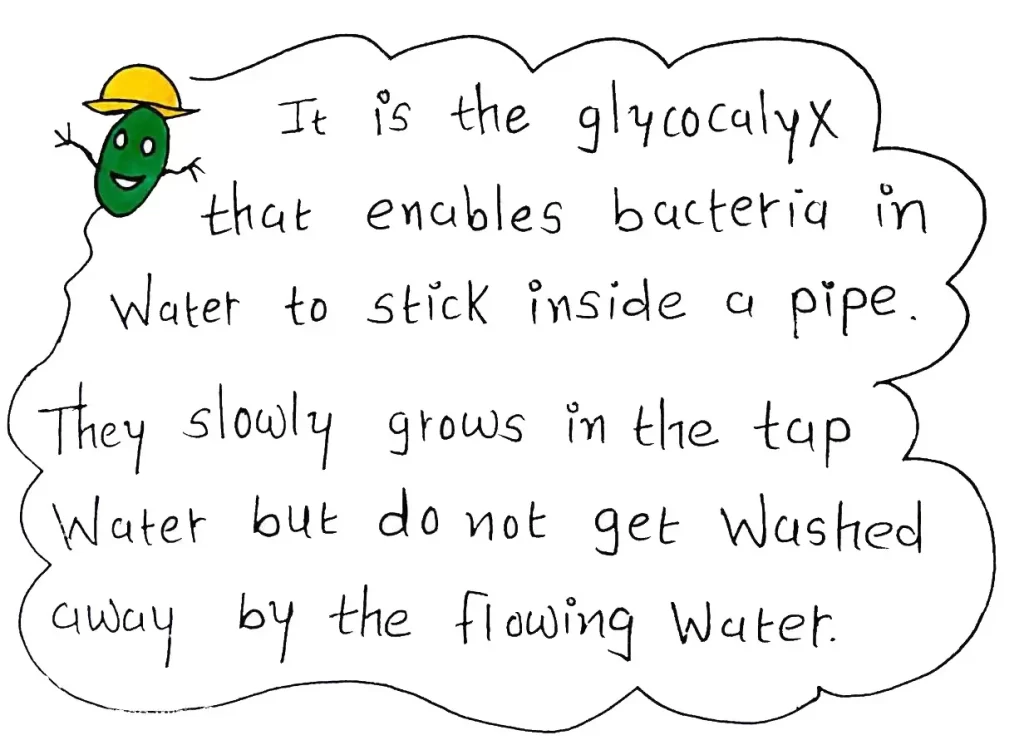
- Through attachment, bacteria can grow on various surfaces like rocks in fast-moving streams, plant roots, human teeth, medical implants, water pipes, etc.
- Example– streptococcus mutans is main cause of caries in teeth, because the bacteria gets attached with teeth surface by glycocalyx.
4.SHEATHS
- Some freshwater and marine water bacterial species form chains or trichomes (outgrowths on plants, algae, lichens etc.) which are enclosed by a hollow tube called sheath.
- Sometimes they may unite with ferric or manganese hydroxides which makes them stronger.
5.PROSTHECEAE AND STALKS
PROSTHECEAE
- These are semi rigid appendages of cell wall and cytoplasmic membrane.
- They are found in number of aerobic bacteria from freshwater and marine water.
- They may be single or several.
- Their main function is to increase the surface area of the cells for nutrient absorption in the dilute environment.
STALK
- They are non-living ribbon like appendages that are extracted from the cell.
- They aid in attachment of the cells to surfaces.
6.THE CELL WALL
- Most of the bacterial cells are having an envelope around cell cytoplasm.
- Cell wall lies below the capsules, sheaths, flagella and above to the cytoplasmic membrane.
- This is a very tough and rigid structure which provide definite shape to the cell.
- As most of the bacteria lives in hypotonic environment, it prevents the cell from swelling and bursting due to water intake.
- It is resistant to extremely high pressure.
- Thickness-10-20 nm.
- Weight-10-40% of the dry weight of bacterial cell.
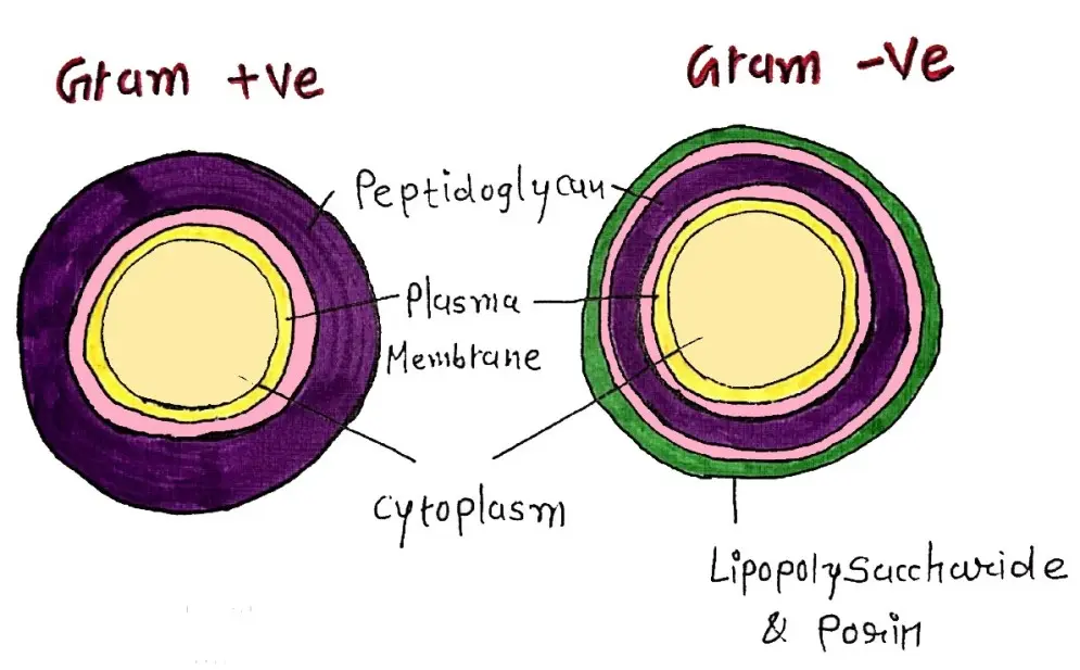
Structure and chemical composition
Made up of number of layers.
The walls of Gram -ve bacteria are generally thinner (10-15 nm) than Gram +ve bacteria (20-25 nm).
Peptidoglycan (murein) is most important component.
- It is made up of 2 polymer and 4 amino acids.
- Polymers are N-acetyl glucosamine (NAG) & N acetylmuramic acid (NAM).
- Amino acids are L-alanine, D-alanine, D-glutamate and diamino acids.
- Two layers of peptidoglycans are cross linked to each other to form a strong framework which provides rigidity to whole body.
- Few antibiotics like penicillin suppresses the synthesis of this framework thus the cell wall synthesis is stopped.
Teichoic acid
- It is a polymer which is negatively charged.
- It is associated with peptidoglycan.
- It forms a major surface antigen.
- It is hydrophilic and it transports positively charged substances to the bacterial cell in the storage of phosphorus.
FUNCTIONS
- Determine the shape of bacterial body.
- It prevents rupture of cell from osmotic pressure.
- It is the important component while cell division.
- It contains so many sites for receptor proteins.
Hope you find this post helpful but wait, it’s not completely done here because we still have to learn about structures that lies inside the bacterial cell wall that will be covered in an another post INTERNAL STRUCTURES OF BACTERIA.





It was really helpful and abundant source of knowledge about one topic .. really helped me to clear my doubts .. whole concept in an understanding language like someone teaching infront of me.. rare gem .
Thank you Priyanka…
Very useful 👌