INTRODUCTION
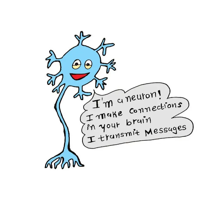
- The neuron is defined as the structural and functional unit of the nervous system.
- It is having nucleus and all the organelles in cytoplasm.
- It is different from other cells in two ways:
- Neuron has branches (processes) called axon and dendrites.
- Neuron does not have centrosome. So, it cannot undergo division.
- Total estimated number of neurons in human brain is more than 1012 .
CLASSIFICATION OF NEURON
Neurons are classified by 3 different methods.
A. Depending upon number of poles
B. Depending upon function
C. Depending upon length of axon.
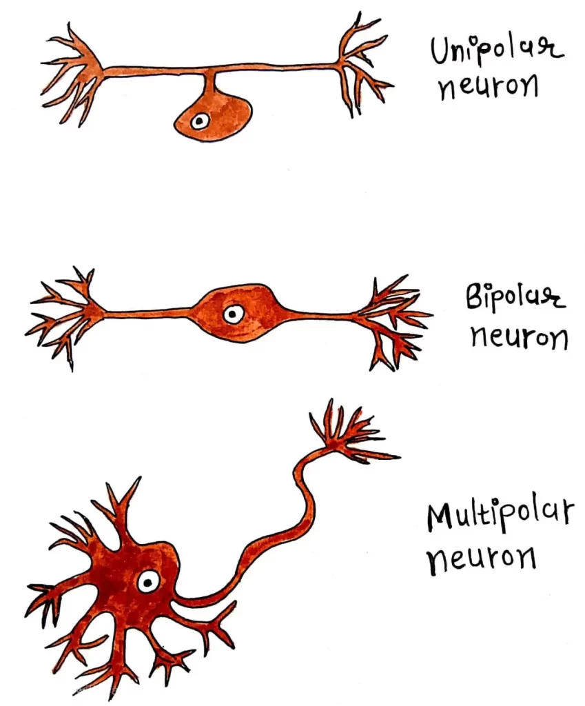
A. DEPENDING UPON THE NUMBER OF POLES
Divided into 3 types:
1. Unipolar neurons
2. Bipolar neurons
3. Multipolar neurons.
1. Unipolar Neurons:
- Have only one pole.
- Both axon and dendrite arise from a single pole.
- Present only in embryonic stage in human beings.
2. Bipolar Neurons:
- Neurons with two poles.
- Axon arises from one pole & dendrites arise from another pole.
3. Multipolar Neurons:
- Have many poles.
- One of the poles gives rise to axon & all other poles give rise to dendrites.

B. DEPENDING UPON THE FUNCTION
Classified into 2 types:
1. Motor neurons (Efferent).
2. Sensory neurons (Afferent).
1. Motor Neurons:
- Carry motor impulses from CNS to peripheral effector organs like muscles, glands, blood vessels, etc.
2. Sensory Neurons:
- Carry sensory impulses from periphery to CNS.
C. DEPENDING UPON THE LENGTH OF AXON
Divided into 2 types:
1. Golgi type I neurons
2. Golgi type II neurons.
1. Golgi Type I Neurons have long axons. Cell body is in different parts of CNS and their axons reach the remote peripheral organs.
2. Golgi Type II Neurons have short axons. present in cerebral cortex and spinal cord.
STRUCTURE
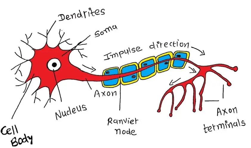
Neuron is made up of 3 parts:
- Nerve cell body
- Dendrite
- Axon.
- Dendrite and axon form processes of neuron.
- Dendrites are short processes and axons are long processes.
- Dendrites and axons are called nerve fibres.

1.NERVE CELL BODY (soma or perikaryon):
- It is irregular in shape.
- It is constituted by cytoplasm called neuroplasm, which is covered by a cell membrane.
- Cytoplasm contains a large nucleus, Nissl bodies, neurofibrils, mitochondria and Golgi apparatus.
- Nucleus: Each neuron has one nucleus, centrally placed which has one or two prominent nucleoli but it does not contain centrosome, cannot multiply like other cells.
- Nissl Bodies: These are small basophilic granules in cytoplasm which are present in soma & dendrite but not in axon & axon hillock.They are responsible for spotted appearance of soma on staining.They contain ribosomes and are concerned with synthesis of proteins.
- Neurofibrils: These are thread-like structures present in the form of network in soma & nerve processes, consist of microfilaments & microtubules.
- Mitochondria: Present in soma & in axon.They form powerhouse of nerve cell, where ATP is produced.
- Golgi Apparatus: Concerned with processing & packing of proteins into granules.
2.DENDRITE:
- They are branched process of neuron & it is branched repeatedly.
- Dendrite may be present or absent.
- If present, it may be one or many in number.
- Dendrite has Nissl granules and neurofibrils.
- They transmit impulses towards the nerve cell body.
- Usually, the dendrite is shorter than axon.
3.AXON:
- Longer process of nerve cell. Each neuron has only one axon.
- Arises from axon hillock & it has no Nissl granules.
- Length of longest axon is about 1 meter.
- Axon transmits impulses away from the nerve cell body.
As now you have the idea about the structure of Neuron, you should also read the Functions of Hypothalamus.
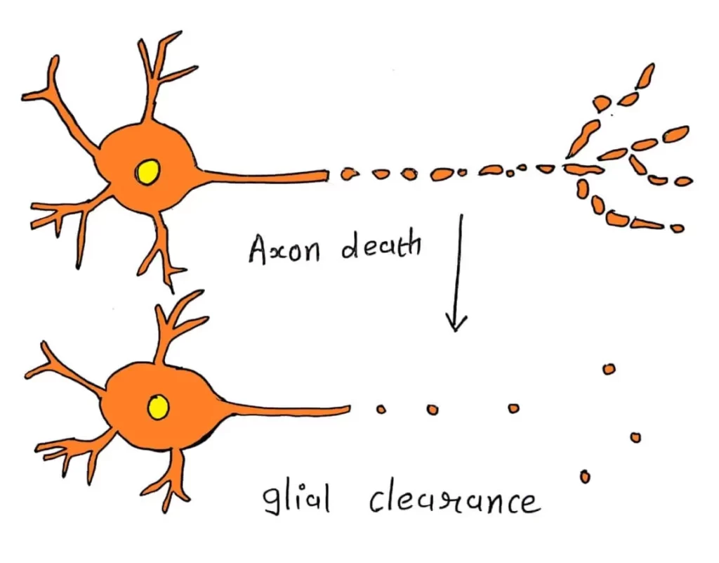
WALLERIAN DEGENERATION
Wallerian degeneration is pathological change that occurs in the distal cut end of nerve fibre (axon).
- It is named after the discoverer Waller.
- It is also called orthograde degeneration.
- Wallerian degeneration starts within 24 hours of injury.

