Cells are connected with one another by intercellular junctions/cell junctions or the junctional complexes.
Table of Contents
ToggleWhere are these junctions located?
Cell junctions are the connection between the neighbouring cells or the contact point between the cell and extracellular matrix. They are also known as membrane junctions.
They are classified into three types
- Occluding junction
- Communicating junction
- Anchoring junctions
Before understanding the types of cell junctions, its important to have some knowledge about the cell adhesion molecules (CAMs).
Read STRUCTURE AND FUNCTIONS OF CELL HERE.
You many also like to read some interesting facts about CELL MEMBRANE by following the links.
CELL ADHESION MOLECULES (CAMs)
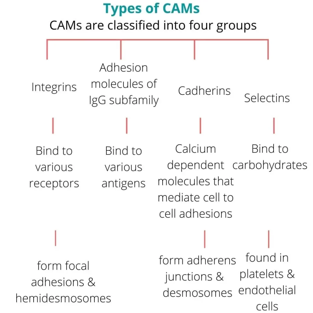
CAMs are different types of proteins that lies on the surface of cell membrane which are also the parts of the intercellular junctions.
Through cell adhesion molecules, cells are attached to each other and also to basal lamina.
TYPES OF CAMs
MECHANISM OF ADHESIONS
The cell adhesion molecules act as binding proteins by 4 mechanisms that are mentioned below
- Anchoring with cytoskeleton- anchors the cytoskeleton inside the cell.
- Homophilic binding- binds to similar molecules of the other cells.
- Heterophilic binding- binds to different molecules of the other cells.
- Binding to laminins– laminins are large cross shaped proteins with many receptors in the extracellular matrix.
Functions of cell adhesion molecules
The main function is to bind the neighboring cells to each other but they also perform some other functions like,
- Transmission of signals inside and out of the cells.
- Anchors tissues together in adults.
- Helps while embryonic development, formation of nervous system and other bodily tissues.
- Plays important role in inflammation and wound healing.
- Acts in metastasis of tumors.
Let’s get back to cellular junctions,
Here is the classification of cell junctions…

OCCLUDING JUNCTIONS
Cell junctions that prevent intercellular exchange of ions and molecules are known as occluding junctions.
e.g., Tight junctions
TIGHT JUNCTION/ ZONA OCCLUDENS/ OCCLUDING ZONE
- They are the intercellular occluding junctions. They prevent the passage of large molecules.
- Structure: a tight junction is made up of a ridge which has two halves (one half from each neighbouring cells).
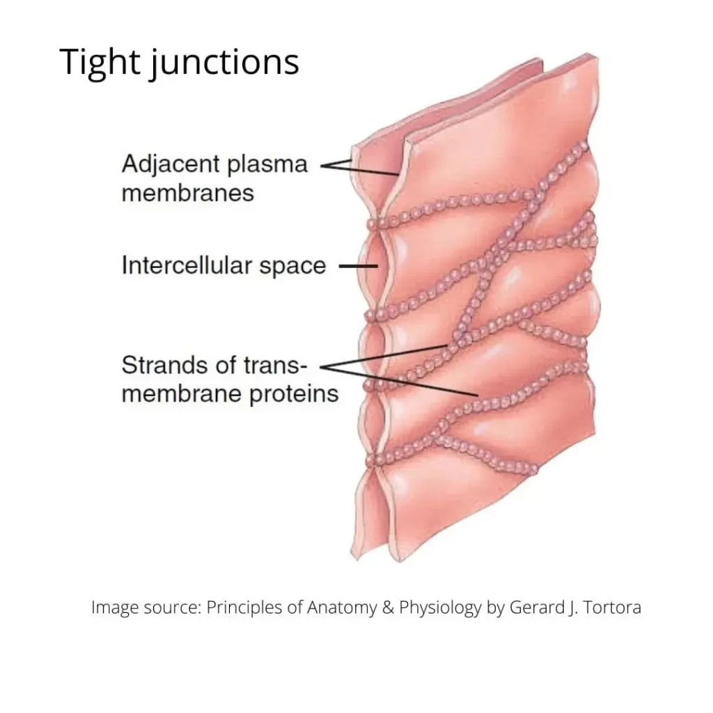
- Both halves of the ridge fuse with each other and create a watertight seal between two adjacent cells.
- At the sight, cells are held tightly against each other by many individual groups of tight junction proteins called claudins.
- They are not randomly located on cell membrane but occupies a precise position at outermost edge of intercellular space so, they are considered a polarized structure.
- The groups are arranged into strands (string) that form a branching network with larger numbers of strands making a tighter seal.
- Purpose: keep liquid from escaping between cells and allowing a layer of cells to act as an impermeable membrane.
- Examples: epithelial cells of stomach, intestine and urinary bladder have many tight junctions that prevent contents of these organs from leaking out into extracellular space.
Functions of tight junction
- Provide strength and stability to tissues.
- Forms a selective barrier for small molecules and a total barrier for large molecules.
- Maintains cell polarity: tight junction acts as a fence and maintains the cell polarity by keeping the proteins in the apical region of the cell membrane.
- Forms blood-brain barrier: tight junctions in brain capillaries forms blood-brain barrier to prevent entry of many substances from capillary blood into brain. Only lipid soluble substances (drugs, steroid hormones) can enter through this barrier.
Clinical physiology related to tight junction
Genetic diseases that alter the proteins of tight junction are:
- Hereditary deafness
- Ichthyosis (dry, thick, scaly skin)
- Sclerosing cholangitis (obstruction of bile duct due to inflammation)
- Hereditary hypomagnesemia (low blood magnesium level)
- Synovial sarcoma (cancer of soft tissues)
Some bacterial and viral infections also can alter the functions of tight junctions.
COMMUNICATING JUNCTIONS
cell junctions that allow the intercellular exchange of ions and molecules are known as communicating junctions.
e.g., Gap junctions, chemical synapse.
GAP JUNCTION/NEXUS
These are the intercellular communicating junctions that allows intercellular exchange of ions and molecules.
Structure:
Gap junction develops when a set of six membrane proteins called CONNEXINS form an elongated donut like structure called CONNEXON.
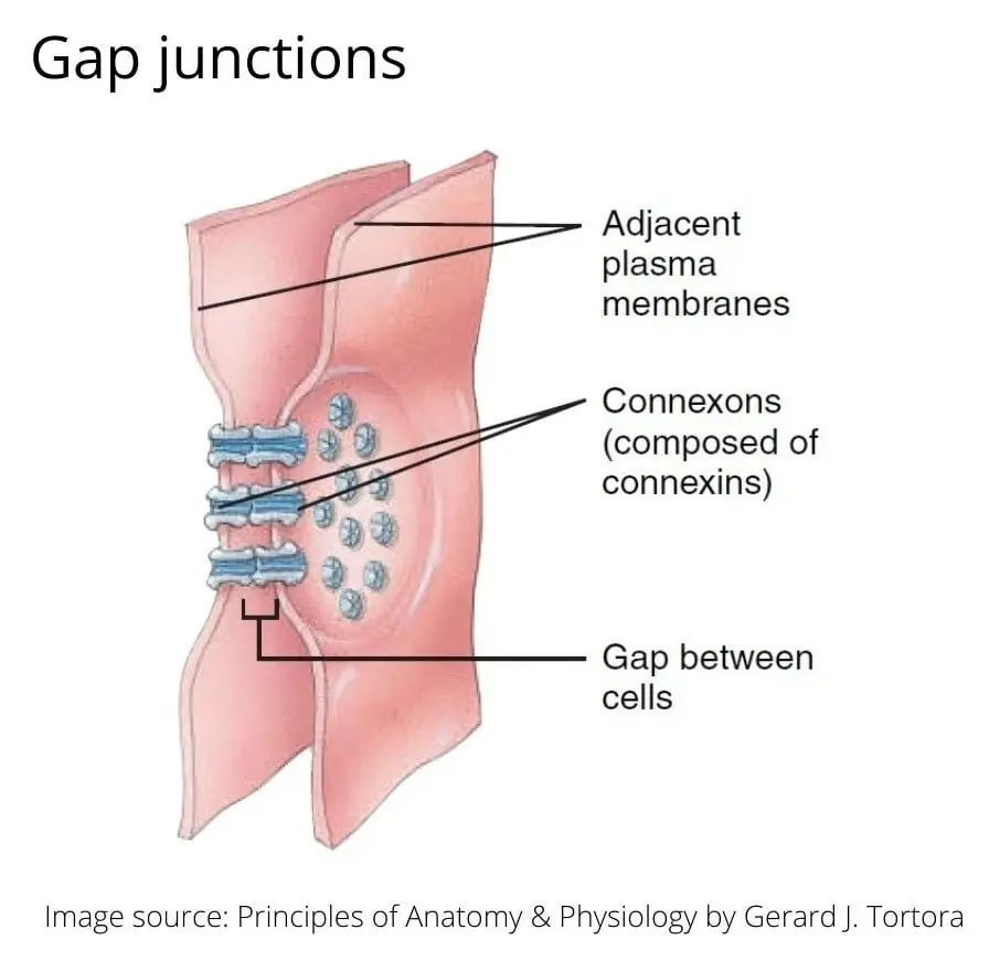
When pores/donut holes of Connexons in neighbouring cells align, a tiny fluid filled channel forms between cells.
These intercellular channels are some 1.5-3nm in diameter.
Through these tunnels, ions and small molecules (molecular weight less than 1000) can diffuse from cytosol of one cell to another cell in neighbour. But large protein molecules that are vital to cells are prevented to pass through it.
Regulation of channel/tunnel diameter in gap junction
Diameter of each channel is maintained by intracellular calcium ions, pH, electrical potential, hormones or neurotransmitters.
Increased concentration of ions causes protein subunits of connexin come close to each other by sliding and so the diameter of channel decreases.
When a cell is injured, gap junction gets closed and isolates a damaged cell from its neighbors. Due to this isolation calcium level gets raised and pH falls in the cytosol of the damaged cell.
Functions of gap junctions
- Allows cells to communicate with one another.
- Transfer of nutrients and wastes takes place through gap junctions in avascular tissues (e.g., lens and cornea of eyes)
- Some chemical and electrical signals travel through gap junctions in developing embryo.
- Rapid spread of chemical or nerve impulse is possible through gap junctions.
E.g., contraction of heart muscles, gastro-intestinal tract and uterus.
CHEMICAL SYNAPSE
Chemical synapses are the intercellular communicating junctions between a nerve fibre to a muscle fibre or between two nerve fibres.
Signals are transmitted by release of chemical transmitters.
Clinical physiology related to communicating junctions
Genetic mutation that alters the connexins can cause diseases like,
- Deafness
- Keratoderma (skin of palm and soles gets thick)
- Cataract (opacity of eye lenses)
- Damage in the nerve cells of peripheral nervous system (peripheral neuropathy)
- Charcot-Marie-Tooth disease (small and weak muscles due to peripheral nerve damage)
- Heterotaxia syndrome (abnormal arrangement of organs of chest and abdomen)
ANCHORING JUNCTIONS
Anchoring junctions provide strong mechanical (structural) attachment between neighbouring cells.
These attachments may be between cell to cell or between cell to extracellular matrix.
They are important for structural integrity of the tissues which are working under severe mechanical stresses.
e.g., Cardiac muscles, epidermis of skin etc.
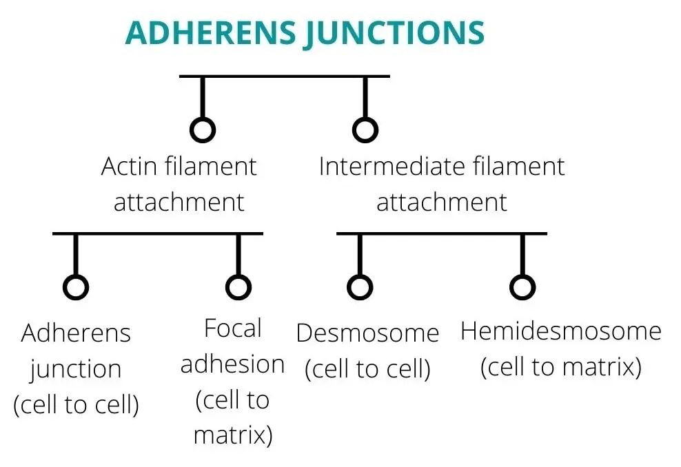
The firm attachment is provided by two types of filaments,
ADHERENS JUNCTION/ZONULA ADHERENS/ADHESION BELTS/BELT DESMOSOMES
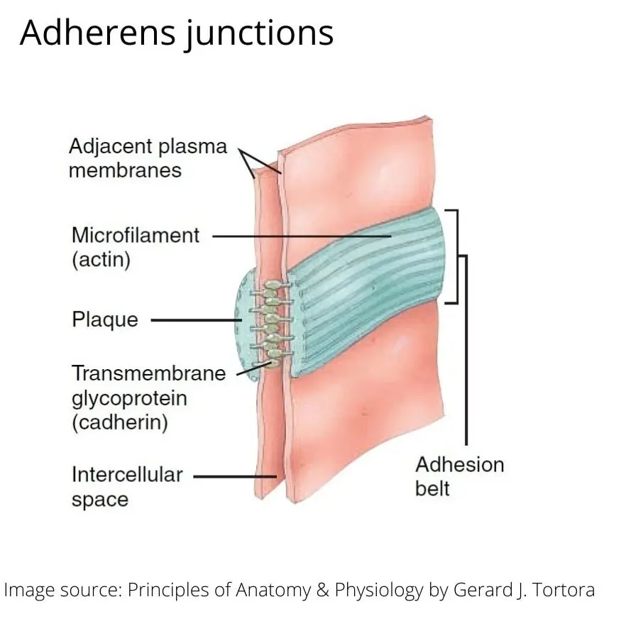
The word meaning of zonula is a small zone or belt like structure. To adhere means to stick together.
They are also known as adhesion belt as they encircle the entire cell just like a belt encircle your waist.
Actin filaments of one cell connects the actin filaments of another adjacent cell.
Adherens junctions are built from CADHERINS (transmembrane proteins) whose intracellular segments bind to CATENINS (a protein that helps in cell adhesion) and extracellular segment bind to each other.
They are located just below the tight junctions.
These provide strong mechanical attachments of neighbouring cells.
FOCAL ADHESION
These are the cell to matrix junctions.
Actin filaments of one cell connects to the extracellular matrix.
The proteins that connect cell to the matrix are called integrins.
In the epithelium of various organs this junction connects cells with their basal lamina.
Clinical physiology of adherens junction and focal adhesions
Dysfunction of these junction in colon causes development of metastatic tumor.
DESMOSOMES/MACULA ADHERENS
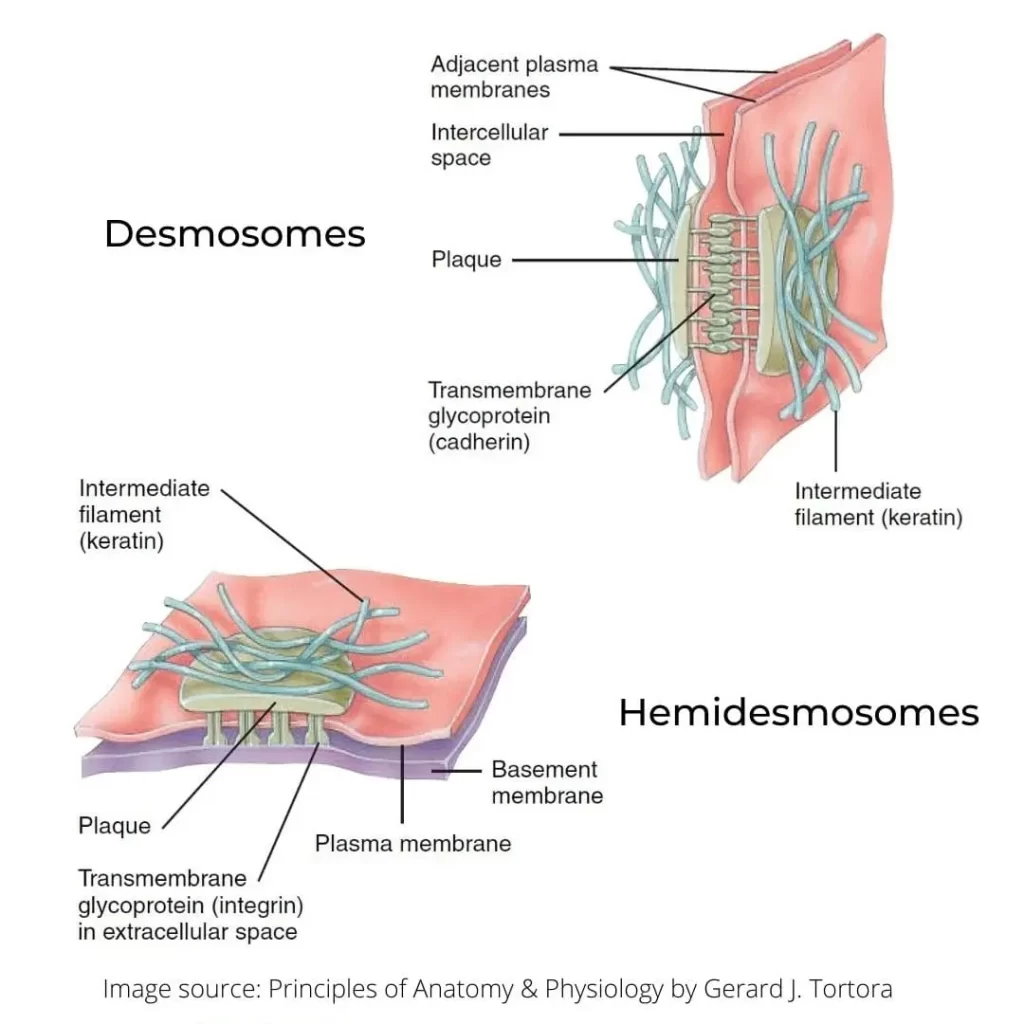
Desmo means a binding body.
Desmosomes are button or spot welds like junctions between neighbouring cells (cell to cell junctions).
These junctions provide strong adhesion between cells.
They form an adhesive bond by connecting to the intermediate filaments of one cell to another cell.
They are randomly located on the lateral sides of cell membrane.
They are found abundantly in the tissues which are subjected to work under continuous mechanical stresses.
e.g., muscles of heart, urinary bladder tissues, mucosa of gastro-intestinal tract.
HEMIDESMOSOMES
Hemi = half
As they look like half of a desmosome they are named as hemidesmosomes.
But the transmembrane glycoproteins are integrins rather than cadherins.
They connect the cells with their basal lamina (to protein laminin).
They do not connect cells with each other but anchor the cells to the basement membrane.
Clinical physiology of desmosomes and hemidesmosomes
Dysfunction causes bullous pemphigoid which is an autoimmune disease with intense blister formation on the skin.
Patient develops antibodies against cadherins due to mutation.




Very well illustrated; one of the small yet important topic made easy to understand 👏🏻👏🏻