They are found in all the natural habitats like soil, water and air.
They form major part of the soil microflora & intestine of animals (E. coli in the intestine of human being).
They can live in all the situations except in pits of volcanoes, deep layers of earth or rock, rain water, distilled water in deep wells, blood of normal animals.
Some species live in extreme hot spring (thermophilic survive on 70°C) while some in extreme cold condition (psychrophilic survive on -190°C).
They can tolerate and remain alive at pH lower than 1 and higher up to 13.
Generally, 1 gm soil contains about 1000-10 million bacteria.
Bacteria is also found in a variety of foods and its products like fruits, vegetables, milk, butter, cheese & milk beverages.
SIZE OF THE BACTERIAL CELL
Bacteria are very small microorganisms which can be visualized only under microscope.
They are stained by staining reagents & then visualized under high magnification power of compound microscope.
They are having size range in microns.
Each bacterial cell measures about 1.25 – 2 µm in diameter & 2-10 µm in length.
Cocci bacteria are about 0.5-2.5 µm in diameter.
Bacilli are 0.3-15 µm in length & 0.2 -2 µm wide.
The smallest rod-shaped Eubacteria is Dialister pneumosintes measure in between 0.15-0.3 µm in size.
The biggest bacteria Beggiatoa mirabilis is about 16-45 µm in diameter and 80 µm in length.
SHAPES AND FORMS OF BACTERIA
Bacterial cells are found in different shapes which are as follows:
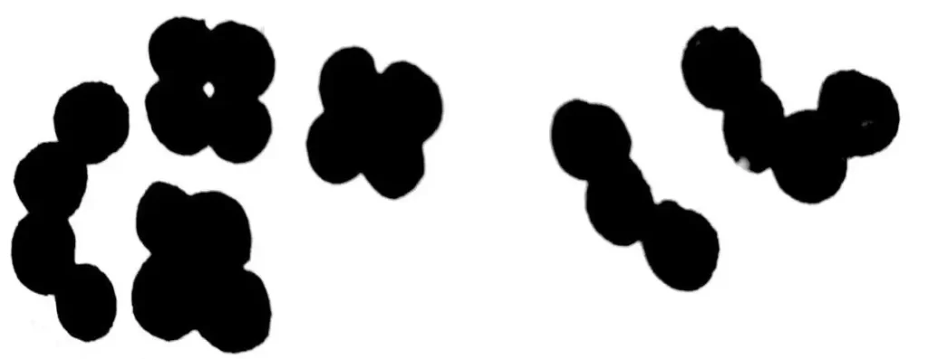
1. Cocci/ Spherical
The Greek word cocci (coccus) means berry.
They are small, simple, oval cells.
They lack flagella.
They are further classified according to their arrangements.
a. Micrococci: Appears singly. e.g. Micrococcus agitis, M. aureus.
b. Diplococci: Pair of cells. e.g. Diplococcus pneumoniae.
C. Streptococci: Rows of cells or in chains e.g. Streptococcus lactis.
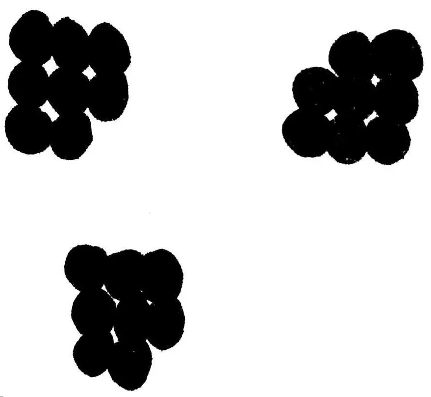
d. Staphylococci: Irregular clusters like bunches of grapes e.g. Staphylococcus aureus, Staphylococcus Albus.
e. Tetra cocci: Group of four e.g. Neisseria and Micrococcus tetrogenus.
f. Sarcinae: produce cuboidal pattern of group of eight cells e.g. Sarcinae lutea.
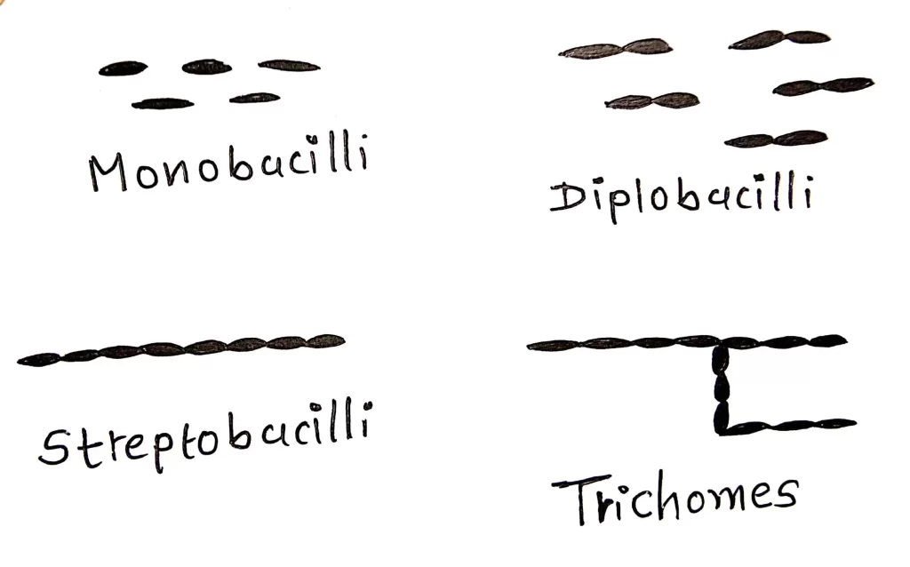
2. Bacilli/ Rod Shaped Bacteria/Bacillus Bacteria
The Greek word bacillus means stick.
They are rod shaped cells.
Their ends may be rounded flat or pointed.
In some of the bacilli the cell length may be equal to the width which are known as coccobacilli.
They may be flagellated or non-flagellated.
Most of the bacteria causing disease in plants are of bacilli type.
They may be of following types:
Monobacillus: Arrange singly.
Diplobacilli: Group of two e.g. Diplobacillus pneumoniae.
Streptobacillus: Appear in chains e.g. Bacillus tuberculosis.
Palisade/ trichomes: Very rarely they arrange in a palisade (fence) arrangement.
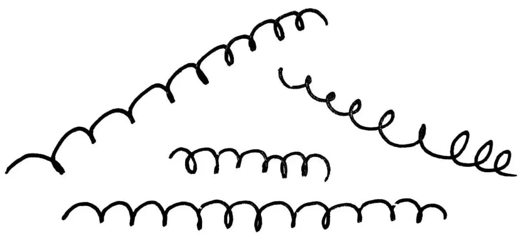
3.Spiral or Helical
The Greek word spira means coiled.
They are long rigid rods with several curves or coils.
They are found as free living, unicellular entity.
They are flagellated e.g. Spirillum minus, S. volutans.
4. Vibrio or Coma
The bacteria of this group are like ‘coma or small curved rod’.
They bear flagella at their end. e.g. Vibrio cholerae.
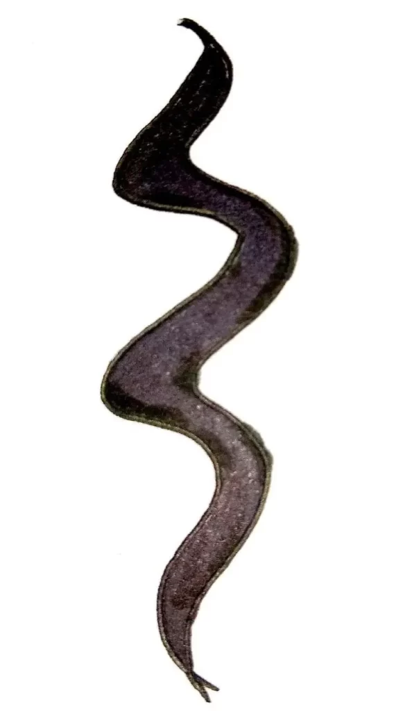
5. Spirochete
These bacteria appear like a cork screw.
Their length is more as compared to their diameter.
Their body is more flexible.
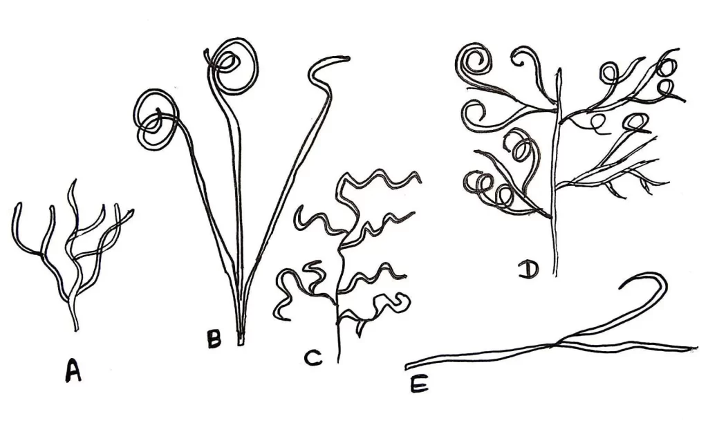
6. Filamentous/ Mycoplasma
They are cell wall deficient bacteria so they do not have stable structure.
They are round or oval bodies with interconnecting filaments.
These types of bacteria are generally found in sewage water and the water coming out from sugar industry.
Ferrous containing water generally contain filamentous bacteria e.g. Nocardia and Beggiatoa.
7. Actinomycetes
Their characteristic shape is because of rigid cell wall.
They are branching filamentous bacteria. e.g. Streptomyces species.
8.Pleomorphic
Many bacteria change their shape & structure according to the change in environmental conditions.
They are found in various forms are known as pleomorphic bacteria e.g. Acetobacter.
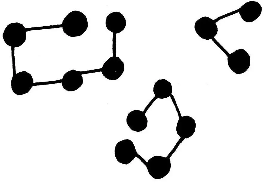
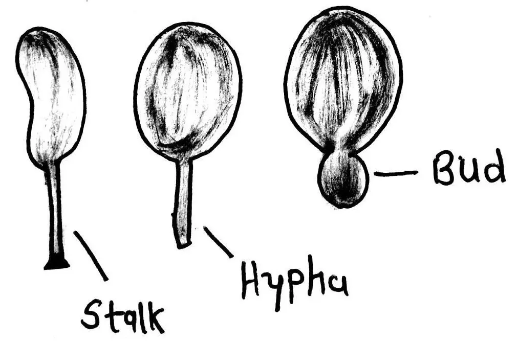
9.Budding Bacteria
These are of football shaped structure with a swollen part & a thin tube.
This tube gradually increases in size & its terminal end swells up to form new cell which is globular and ultimately a cell network is formed e.g. Rhodomicrobium.
ULTRASTRUCTURE OF BACTERIAL CELL
As the bacteria is very small in size, an electron microscope is used for clear visualization of internal bacterial structure.


Of course, what a great site and informative posts, I will add backlink bookmark this site? Regards, Reader.