In this post, we will study about Gram Staining Which is most widely used staining method in Microbiology.
INTRODUCTION
- Bacteria are colourless so their structural details cannot be seen under light microscopy due to lack to contrast.
- So, it is necessary to produce colour contrast to improve visibility by using staining methods.
- There are many staining methods used for different purposes like Simple stain, Negative staining, Impregnation methods, gram stain, acid-fast stain, albert stain etc.
[Note: Words with ( * ) are particularly defined at the end of this post.]
COMMON STAINING TECHNIQUES USED IN MICROBIOLOGY
- Simple stain
They give the colour contrast, but diffuse the same colour to all the bacteria in a smear.
Basic dyes, like methylene blue or basic fuchsin are the examples of simple stains. - Negative staining
A drop of suspension of microbes is mixed with dyes so, the background gets stained black while unstained bacterial/yeast capsule project in contrast.
This technique is very useful in the examination of bacterial/yeast capsules which do not absorb simple stains.
Examples are India ink or nigrosine. - Impregnation methods
Impregnation means come into contact with or to absorb something.
Microorganisms which are very thin to be seen under the light microscope, are thickened by impregnation of silver salts on their surface to make them visible.
Example: Demonstration of bacterial flagella and spirochetes. - Differential stain
Here, two stains are used which can reveal different colours of different bacteria or bacterial structures, which help to differentiate bacteria.
The most commonly used differential stains are:
• Gram stain: Used to differentiate bacteria into gram positive and gram-negative groups.
• Acid-fast stain: Used to differentiate bacteria into acid-fast and non-acid-fast groups.
• Albert stain: Used to differentiate bacteria having metachromatic granules from other bacteria which do not have them.
Table of Contents
ToggleGRAM STAINING METHOD
- Gram staining is the common procedure of staining (colouring) Bacteria.
- It was developed by Hans Christian Gram (1884).
- This method is used to differentiate two large groups of bacteria on the basis of their different cell wall constituents.
- Gram-positive bacteria
- Gram-negative bacteria.
- This stain also allows the clinician to determine whether the organism is round or rod-shaped.
- For any stain you first need to smear the substance to be stained (sputum, pus, etc.) onto a slide and then heat it to fix the bacteria on the slide.
4 steps of the Gram staining procedure
STEP-1: CRYSTAL VIOLET
- Pour on crystal violet stain (a blue dye) and wait for 1 minute.
- It stains them all the same purple colour.
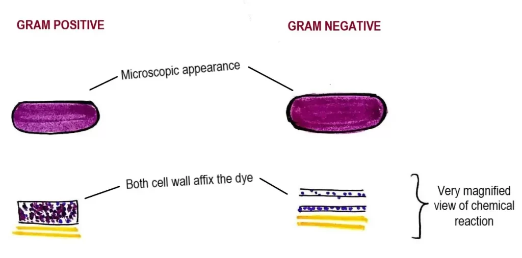
STEP-2: GRAM’S IODINE
- Wash off with water and flood with Gram’s iodine solution & wait for 1 minute.
- This is a stabilizer that causes the dye to form large complexes in the peptidoglycan meshwork of the cell wall.
- These dye complexes are easily trapped in the thicker gram-positive cell walls as compared to gram negative cell walls.
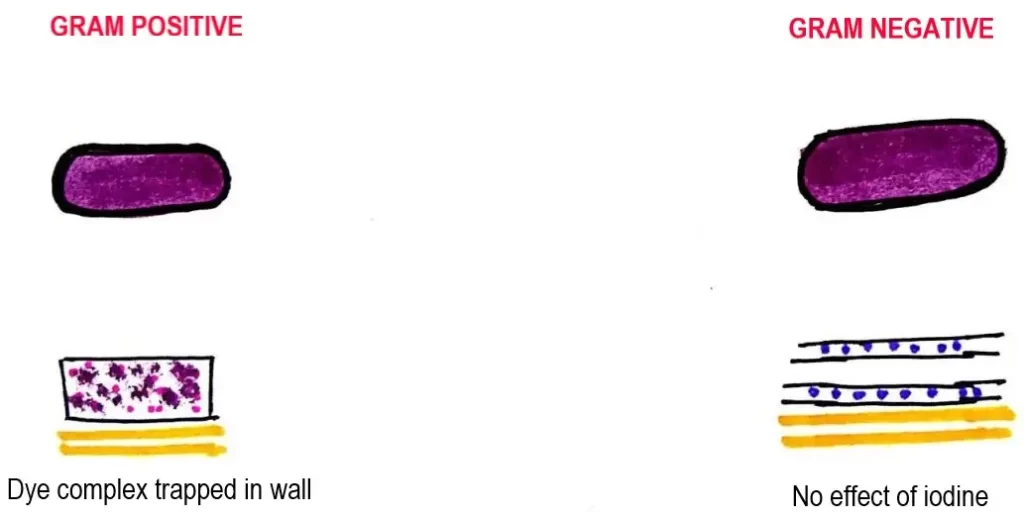
STEP-3: DECOLOURIZATION
- Wash off with water & then “Decolorize” with 95% ethyl alcohol for 20-30 seconds.
- Alcohol dissolves lipids in the outer membrane & removes the dye from peptidoglycan layer-happens only in the gram-negative cells.
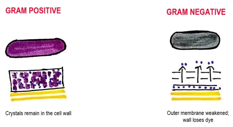
STEP-4: SAFRANIN (RED DYE)
- Finally, counter-stain with safranin (a red dye).
- Wait 30 seconds and wash off with water.
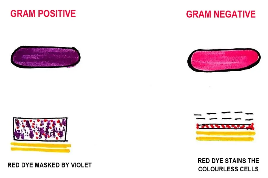
- As gram-negative bacteria become colourless after decolourization, applying the safranin in final step stains the colourless cell red.
INTERPRETATION
Smear is examined under oil immersion objective lens.
Gram-positive resist decolourization & retain the colour of primary stain i.e. VIOLET.
While, if the crystal violet is washed off by the alcohol, these cells will absorb the safranin and appear red. These are called Gram-negative organisms.
Gram-positive = VIOLET
Gram-negative = RED
The different stains are the result of differences in the Cell walls of Gram-positive and Gram-negative bacteria.
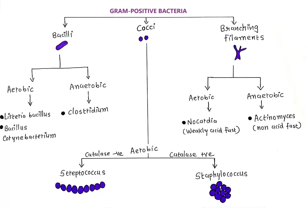
GRAM POSITIVE BACTERIA
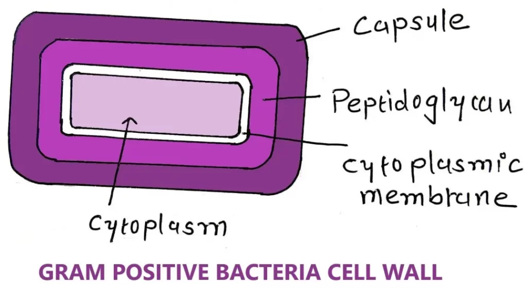
- They retain the colour of crystal violet and stain purple or dark blue.
- During staining procedure, they resist decolorization with absolute alcohol.
Cell wall
- Single layered.
- Straight & even.
- Less elastic & more rigid.
- This rigidity is due to the high amount of peptidoglycan (80%).
- Cell wall has muramic acid (16-20% of dry weight of cell).
- Resistant to alkaline medium.
- Teichoic acid is present in many bacteria.
- Cell wall is 20-30 nm thick. (sometimes can be 80 nm).
Other characteristics
- Absence of outer layer of lipopolysaccharide.
- Low content of lipid & lipoproteins (1-4%).
- Absence of porins*.
- Periplasmic space absent or very narrow.
- During unfavourable conditions they produce endospores.
- Produces exotoxins normally.
- They show high tolerance towards dryness.
EFFECTS OF ANTIBIOTICS
- Vancomycin antibiotic is used to kill them.
- Cell shows less susceptibility towards Chloramphenicol, Streptomycin & Tetracyclines.
- Cell shows high susceptibility towards Sulphonamide & Penicillin.
Protoplast
If cell wall is removed from the Gram-positive bacteria then they are called as protoplast.
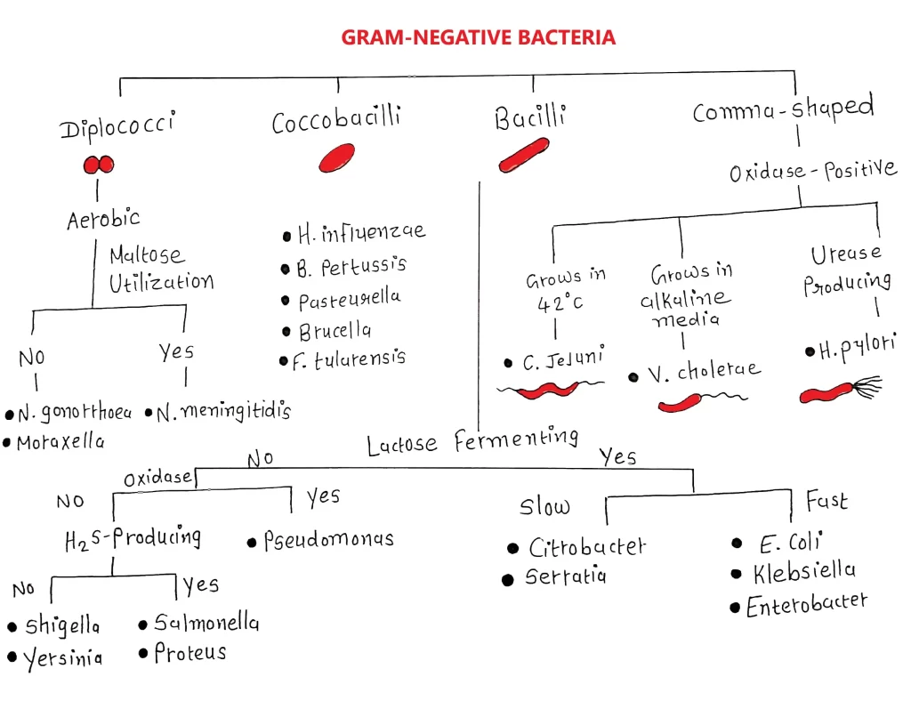
GRAM NEGATIVE BACTERIA
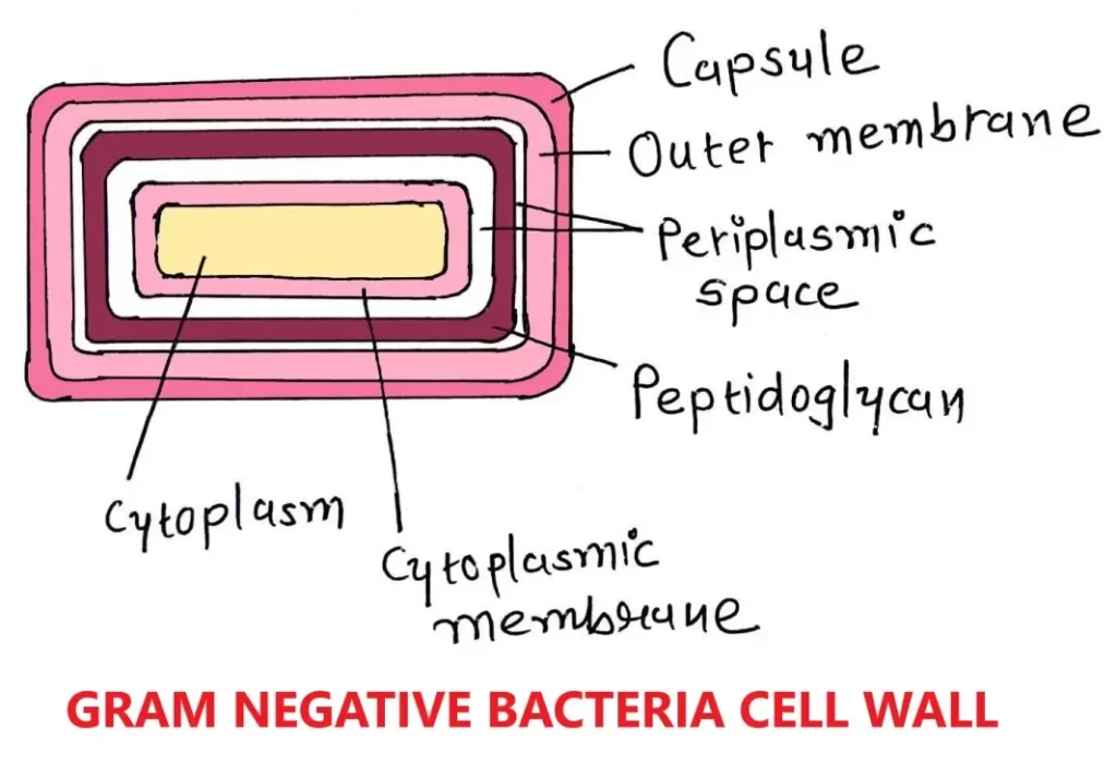
- They are not able to retain crystal violet colour.
- During staining procedure, absolute alcohol dissolves an outer membrane.
Cell wall
- Wavy & uneven.
- More elastic & less rigid.
- This elasticity is due to less amount of peptidoglycan (2-12%).
- Cell wall has less muramic acid (2-5% of dry weight of cell).
- Sensitive to alkaline medium.
- Teichoic acid is absent.
- Cell wall is 8-12 nm thick.
Other characteristics
- Presence of outer layer of lipopolysaccharide.
- High content of lipid & lipoproteins (11-22%).
- Presence of porins.
- Periplasmic space present.
- They do not produce endospores.
- Produces endotoxins normally.
EFFECTS OF ANTIBIOTICS
- Vancomycin antibiotic is not effective.
- Cell shows less susceptibility towards Chloramphenicol, Streptomycin & Tetracyclines.
- Cell shows high susceptibility towards Sulphonamide & Penicillin.
SIMILARITIES
- Both are prokaryotic organisms.
- They do not have well developed nucleus, nuclear membrane & other membrane-bound organelles.
- Their cell membrane is made up of peptidoglycan.
- Their cell cytoplasm is surrounded by the lipid bilayer.
- They have closed circular DNA as a genetic material.
- They both contain s-layer on their surfaces.
- They both goes under Sexual and Asexual reproduction.
Sexual reproduction*- Transformation & Transduction.
Asexual reproduction*- Binary fusion.
- Both have many flagellated & non-flagellated species.
REFERENCE TERMINOLOGIES
Asexual reproduction– Production of living organism with no fusion of gametes or change in chromosome numbers. Includes fission, fragmentation, budding, vegetative reproduction, spore formation & agamogenesis.
Porins– Membrane channels made up of proteins.
Sexual reproduction– Production of new living organisms by combination of two different types of genetic information from two different individuals.

Very well explained , thank you for making it easy for understanding