As we already know that the cellular organelles are permanent components of the cells, this post focuses on the membrane bound organelles of the cell.
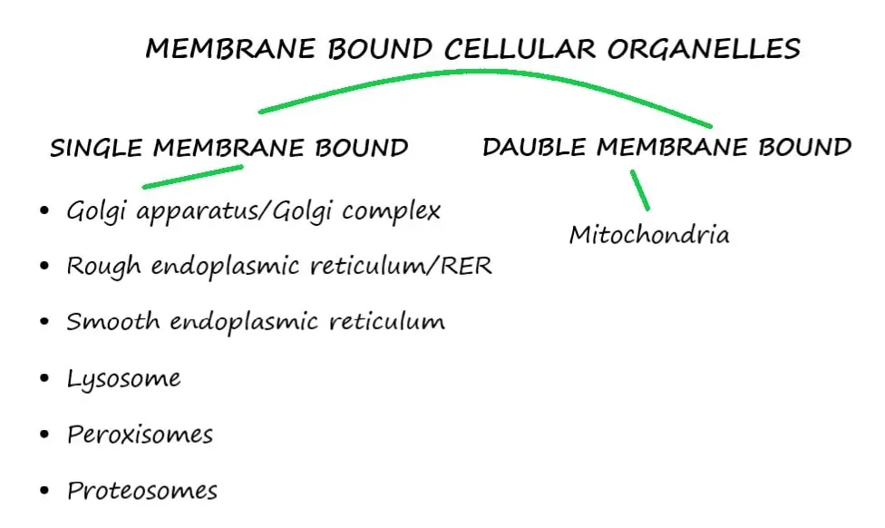
You can revise the classification of the organelles in below given image.
You can read about non-membrane bound cellular organelles by following the link here,
NON-MEMBRANE BOUND CYTOPLASMIC ORGANELLES.
Table of Contents
ToggleSingle membrane bound organelles are,
- Golgi apparatus/Golgi complex
- Rough endoplasmic reticulum/RER
- Smooth endoplasmic reticulum/SER
- Lysosome
- Peroxisomes
- Proteosomes
Double membrane bound cytoplasmic organelle is mitochondria which is described in a separate post here, Mitochondria/ The power house of the cell.
GOLGI APPARATUS/GOLGI COMPLEX
This is a membrane bound cellular organelle.
Named after its founder Camilo Golgi.
It is a collection of 2 to 20 vesicles/ sacs/ tubules which are known as cisternae.
Usually, each cell has only one Golgi complex but some may have more than one.
Red blood cells do not contain Golgi bodies.
It deals with the processing of proteins.
Structure of Golgi Apparatus
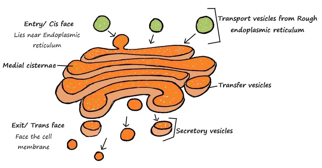
Situated near the nucleus.
Space between entry and exit faces are called medial cisternae.
Curved cisternae give the cuplike shape of Golgi complex.
The opposite ends of Golgi complex are different in size, shape, content and enzymatic activities.
Entry face: processes the materials. Receives proteins from Rough ER and modifies them.
Medial cisternae: forms glycoproteins by adding carbohydrate to proteins and forms lipoproteins by adding lipids to proteins.
Exit face: further modifies the protein molecules. Sorts and packages them for transport to their destination.
Functions of Golgi Apparatus
Processing, packaging, labelling and delivery of proteins, lipids, carbohydrates and other molecules to the different parts of the cell.
Processing: vesicles containing proteins are processed and modified into glycoproteins and lipoproteins.
Packaging: all the processed molecules are packaged in the form of lysosomes, secretory granules and secretory vesicles.
Also called as “Post office of the cell”.
ENDOPLASMIC RETICULUM/ER
- Plasmic= cytoplasm
- Reticulum= network
The Endoplasmic reticulum is a membranous network of interconnected flattened tubules.
It is connected to nuclear outer membrane and projects all over the cell cytoplasm.
Types of endoplasmic reticulum
Morphologically two types of ER have been identified. Both are interconnected and continuous with one another. Both are concerned with different types of function.
- Rough endoplasmic reticulum
- Smooth endoplasmic reticulum
- Rough endoplasmic reticulum/Granular endoplasmic reticulum
- Continuous with nuclear outer membrane.
- Lies folded into interconnected flat tubules or vesicles.
- Ribosomes attached to its outer surface is responsible for the rough appearance (causing rough or bumpy look).
Functions of Rough ER
Protein synthesis
Ribosomes attached to granular endoplasmic reticulum arrange the amino acid chains into small protein units and transport them into Rough ER.
These protein units inside the ER are further processed and sorted.
Here, some enzymes add carbohydrates into protein units to form glycoproteins.
While other enzymes add phospholipids into protein units.
Cells which are active in protein synthesis, develop large number of Rough ER
e.g., Acinar cells of pancreas, Nissl granules of nerve cells, Russell’s bodies of plasma cells.
- Break down of exhausted organelles
Rough endoplasmic reticulum wraps itself around exhausted organelles and forms a vacuole in which this they are digested by lysosomal enzymes; this process is known as autophagy.
- Smooth endoplasmic reticulum/ Agranular endoplasmic reticulum/ Tubular endoplasmic reticulum
Extends from Rough endoplasmic reticulum.
Formed by a network of many connected tubules.
Does not have Ribosomes attached to its outer surface (causes smooth appearance).
Functions of Smooth ER
It is functionally more diverse than Rough ER due to presence of unique enzymes.
It does not synthesize proteins as it does not have ribosomes.
- Synthesis of non-protein substances
They produce fatty acids and steroids like estrogen and testosterone.
Well developed in Leydig cells and cells of adrenal cortex.
- Role in various metabolic processes
Outer surface of smooth endoplasmic reticulum contains many enzymes that play an important role in cellular metabolism.
e.g., Smooth ER enzymes in the cells of liver, kidneys and intestine separates the phosphate group from glucose-6-phosphate which allows entry of free glucose molecules into bloodstream.
- Storage and metabolism of calcium ions
Smooth ER is a major site of storage and metabolism of calcium ions (Ca⁺).
In skeletal muscle cells, calcium ions are released which is important to trigger the muscular contraction.
- Catabolism and detoxification
Enzymes of Smooth ER inactivates or detoxifies the lipid soluble or harmful drug substances.
Applied physiology
- Patients who are habitually taking certain drugs like phenobarbital, may develop changes in smooth endoplasmic reticulum of their liver cells.
- Prolonged use of these drugs causes increase in drug tolerance.
- Because smooth endoplasmic reticulum and its enzymes gets increase in number and quantity in order to protect the cell from the toxic effects of drugs.
- Which means higher and higher doses of drugs are required to achieve the desirable effects.
- This may result in increased chances of drug dependence or over dose.
LYSOSOME
- Lyso= dissolving
- Somes= bodies
Also known as Garbage system of the cell due to their function of degradation.
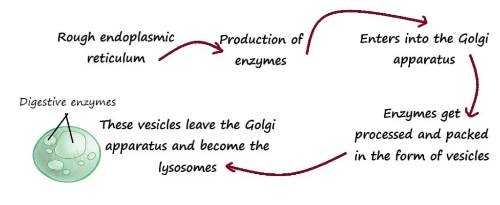
These are round or oval shaped membrane bound organelles which are formed from Golgi apparatus.
Found throughout the cell cytoplasm.
Lysosomal membrane
As lysosomes can contain about 60 different kinds of powerful digestive or hydrolytic enzymes, they have the thickest covering membrane among the cytoplasmic organelles.
The membrane is made up by double layer of lipids.
As the acidic environment is best for lysosomal enzymes to work, the internal acidic pH of 5 (which is hundred times more acidic than cytosol pH which is 7) is needed to maintain.
So, lysosomal membrane contains active transport pumps to import Hydrogen ions (H⁺).
Transporters that can move final digested products to cytosol (like glucose, amino acids, fatty acids) are also contained by lysosomal membrane.
Types of lysosomes
Mainly 3 types are identified
1. Primary lysosomes/ storage vacuoles
They are formed by Rough ER from various hydrolytic enzymes.
Processed and packed in Golgi complex.
They are inactive forms even though having hydrolytic enzymes.
2. Secondary lysosomes/ autophagic vacuoles
Fusion of primary lysosomes with damaged or worn-out cellular components form the secondary lysosomes.

They are active lysosomes.
3. Residual bodies
These are undigested materials in the lysosomes.
Examples of important lysosomal enzymes
Proteases, lipases, amylases, nucleases etc.
Functions of lysosomes
- Lysosomes digest the substances that enter into the cell via endocytosis and then transport back the final digested products into the cytosol.
- Lysosomes can digest the worn-out organelles by the process of autophagy. Also causes the deterioration of tissues that happen soon after death.
- Fulfill extracellular digestion
Example: the head of sperm cell releases lysosomal enzymes for acrosomal reaction.
So, with the powerful digestive and hydrolytic enzymes, lysosome can act within as well as outside of the cell.
Applied physiology
There are some diseases which are caused by defective or absent lysosomal enzymes.
e.g., Tay -sacs disease (Ta-Sacs)
- It is an inherited condition of absent lysosomal enzyme Hex A.
- This Hex A enzyme digests a membrane glycolipid (ganglioside GM2) which is abundant in nerve cells.
- With the absence of Hex A enzyme ganglioside GM2 accumulates in nerve cells.
- As a result, nerve cells cannot function efficiently.
- Mainly affects the children of Ashkenazi (Jewish of east Europe).
- Patient experiences seizures and rigidity of muscles.
- Child gradually becomes blind, lunatic or clumsy.
- They usually die before the 5 years of age.
- With the advancement of genetic science, the adult carrier having defective gene can be detected.
PEROXISOMES
- Peroxi= peroxide
- Somes= bodies
These are single membrane bound organelles.
Spherical in shape.
Structure similar to lysosome but smaller in size.
Contains many oxidases.
Oxidases are the enzymes that can remove hydrogen atoms from various organic substances (we simply can say they can oxidize).
During process of metabolism, along with oxidation of amino acids and lipids, some toxic substances (like alcohol) also get oxidized in peroxisomes.
This is the reason peroxisomes are found in abundance in liver cells where detoxification of many harmful drug substances and alcohol takes place.
But as a result of oxidation reactions, formation of some by-products like Hydrogen peroxide (H₂O₂) and free radicals like superoxide also takes place.
If these metabolic byproducts get accumulated inside of the cell, they can cause the cellular death due to their toxic compounds.
So, peroxisomes have solution for them, they contain an enzyme that can break down H₂O₂ which is known as enzyme catalase.
Also contains destroying enzymes for superoxide.
In this way, production and degeneration of H₂O₂ takes place in the same peroxisome. So, cell gets protected from toxic by-products.
They can self-replicate that means a new peroxisome can arise from prior ones. The pre-existed peroxisomes get enlarged and then divide.
Peroxisomes are the major site for Beta-oxidation of fatty acids.
Functions of Peroxisomes
- Break down of fatty acids
- Detoxification of hydrogen peroxide and other metabolic by-products
- Utilization of oxygen
- Promotion of gluconeogenesis
- Degrades purines to uric acids
- Participate in myelin formation
- Plays a role in bile acids formation
PROTEOSOMES
The word meaning of proteosomes is protein bodies.
Structure
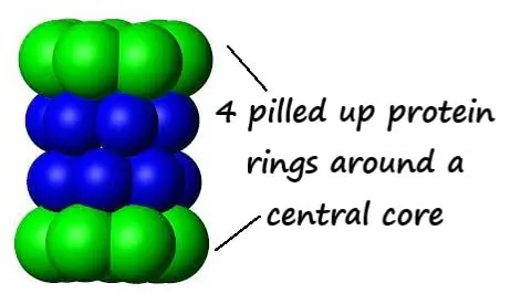
These are tiny barrel shaped structures contains 4 pilled up protein rings around a central core.
Present in both cell cytosol and nucleus.
There are thousands of proteosomes in a typical cell.
They contain proteolytic enzymes. Enzymes that break proteins into small peptides.
As you know that in cell, proteins delivered in the form of vesicles are metabolized in lysosomes.
As well, protein molecules present in cytoplasm also needs to be digested time to time.
So, continuous degeneration of non-required, damaged or defective proteins are done in proteosomes by cutting them into small peptides.
Function of Proteosomes
Degenerates nonrequired, damaged or defective proteins by cutting them into small peptides.
Applied physiology
Accumulation of non-functional misfolded proteins in the cells of brain may result due to failure of proteosomes to degenerate abnormal proteins.
Happens in patients with Creutzfeldt-Jakob disease, Alzheimer’s disease, Parkinson’s disease, amyloidosis and a wide range of other disorders.
Read the Non membrane bound cellular organelles here… CELLULAR ORGANELLES: STRUCTURE AND FUNCTIONS.
The double membrane bound organelle is available here… MITOCHONDRIA/THE POWER HOUSE OF CELL.
You can also read the STRUCTURE AND FUNCTIONS OF CELL and an interesting post on CELL MEMBRANE by following the links.
